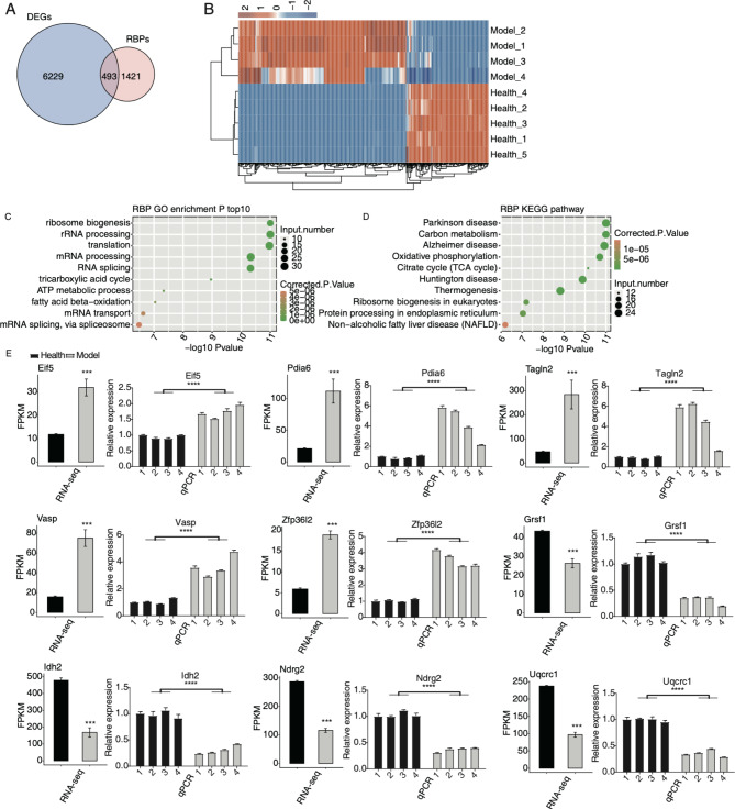
Fig. 2.
Analysis of the differential expression of RBPs in MIRI. (A) The overlapped genes between DEGs and RBPs are shown in venn diagram. (B) The expression levels of the DE RBPs are shown in hierarchical clustering heatmap. (C) The scatter plot shows the top ten GO BP results of the DE RBPs. (D) The scatter plot shows the top ten KEGG pathways of the DE RBPs. (E) Bar plot shows the statistical differences and expression patterns of the DEGs of some critical RBPs. Mean ± SEM is represented by error bars. Student’s t-test; *** P-value < 0.001; **** P-value < 0.0001

