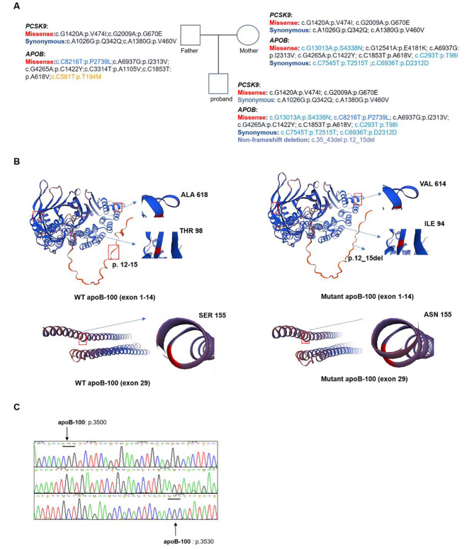
Fig. 4.
Summary of gene variants in APOB and PCSK9. (A) Based on the results of WGS, the mutations in the APOB and PCSK9 genes for the proband and the parents were compiled. (B) SWISS-MODEL was employed to generate a structure depicting the variants located in APOB (Exon 1, exon 4, exon 14 and exon 29). Variants in exon1 and exon 29 led to a change in the spatial conformation of the protein. (C) Sanger sequencing confirmed that the proband had no nucleotide variants at the pathogenic mutation sites prone to occur in the apoB-100 protein (p.3500–3530)

