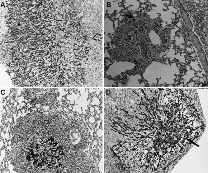
FIG. 6.
Histopathology of the lungs of mice 6 days after infection with 107 A. fumigatus. Hematoxylin and eosin stain combined with Gomori’s methenamine silver was used. Three mice were examined for each treatment group. Magnifications, × 400. (A) Untreated control mice. Large numbers of hyphae are seen invading the bronchioles. Foci of hyphae are surrounded by a modest infiltrate of predominantly monocytic cells with a few PMNs (open arrow). (B) Mice treated with SCH56592 at 10 mg/kg/day. Few hyphae are seen (solid arrow), with modest surrounding infiltrates of mononuclear cells. (C) G-CSF at 600 μg/kg/day. Many large collections of PMNs and necrotic debris (open arrow) surround foci with large numbers of hyphae present. (D) G-CSF and SCH56592. Large abscesses with PMNs surrounding foci of A. fumigatus hyphae. Hyphae (solid arrow) are more numerous than in mice treated with SCH56592 alone.

