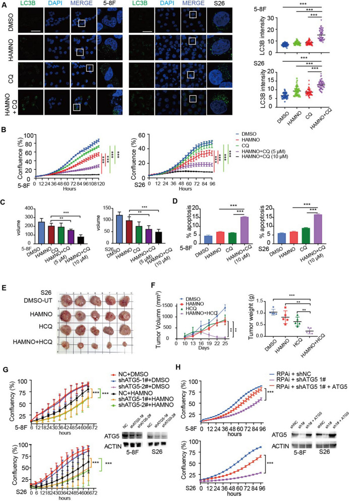
Fig. 5.
RPA inhibition enhanced the sensitivity of NPCs to hydroxychloroquine-mediated inhibition of autophagy. a Quantitative result of LC3B puncta in 5-8F and S26 cells following treatment with HAMNO (5 µM) in the absence or presence of CQ (10 µM) for 48 h. Scale bar = 50 μm. b Confluence of 5-8F and S26 cells treated with HAMNO (5 µM) alone or in combination with CQ (5 µM or 10 µM) was analyzed by the IncuCyte system. c Tumoroids treated with HAMNO (5 µM) alone or in combination with CQ (5 µM or 10 µM) were analyzed by the IncuCyte system. The volume was measured by ImageJ. The mean of five independent experiments is plotted. d The percentage of apoptotic cells induced by HAMNO or CQ alone or in combination for 48 h was determined via Annexin V/7-AAD staining and flow cytometry. The histogram represents the quantification of three independent experiments. Data are the mean of three independent experiments. e Images display tumor size at the end of treatment. f Xenograft tumor growth curve of S26 cells treated with HAMNO (1 mg/kg) or HCQ (60 mg/kg) alone or in combination (left). The scatter plot displays the tumor weight at the end of treatment (right). Error bars indicate the mean ± SEM. for n = 5 biologically independent animals. g Growth curves of ATG5 knockdown NPC cells treated with RPAi. h Cell growth curve of shATG5 knockdown-NPC cells transfected with pcDNA3.0-ATG5 overexpression plasmid or an empty vector control

