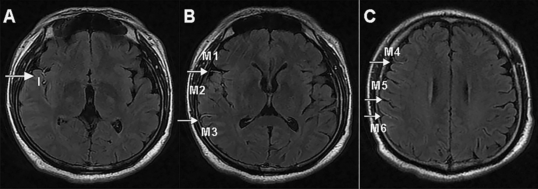
Fig. 1.

Images illustrating FVH: blood supply in area of the left MCA (M1, M2, M3, Insular, M4, M5, M6) showing FVH and FVH-ASPECTSTS score was 7; I, insular cortex; M1, the anterior MCA cortex, corresponded with the frontal operculum; M2, the MCA cortex lateral to the insular ribbon, corresponded with the anterior temporal lobe; M3, the posterior MCA cortex, corresponded with the posterior temporal lobe. M4, M5, and M6, the anterior, lateral, and posterior MCA territories immediately superior to M1, M2, and M3, respectively
