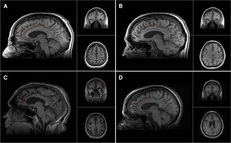
Figure 1.
Cingulate and paracingulate sulci identification and measurement. A 58-year-old male with probable bvFTD displays a leftward pattern of paracingulate asymmetry, A and B. A. The left hemisphere displaying a traced ‘present’ (length ≥ 20 mm), left paracingulate sulcus (red) and a traced cingulate sulcus (yellow). B. The right hemisphere displays a traced cingulate sulcus (yellow) with absence of a right PCS. C. A 62-year-old male with definite bvFTD, images display a traced ‘present’ right paracingulate sulcus, 23 mm in length (red). D. A 60-year-old female with probable bvFTD, images display a traced ‘prominent’ (length ≥ 40 mm) right paracingulate sulcus, 53 mm in length (red).

