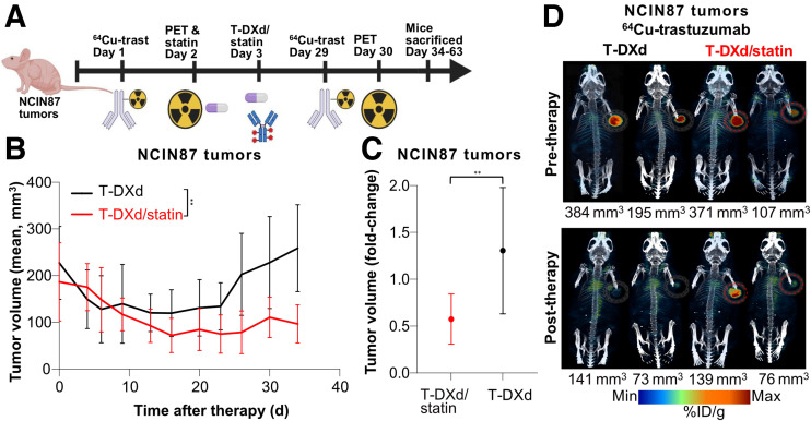
FIGURE 3.
(A) Schematic illustrating T-DXd and statin dose administration across single-dose regime. NCIN87 gastric xenografts were established in female nude mice (9 per group). Once tumors reached about 200 mm3, mice were intravenously administered T-DXd, 5 mg/kg weekly (for 1 wk—single-treatment regime). Lovastatin (4.15 mg/kg) was orally administrated 12 h before and simultaneously with intravenous injection of T-DXd. [64Cu]Cu-NOTA-trastuzumab was intravenously administered on days 1 and 29, and PET images were collected before (day 2) and after (day 30) therapy. Mice were sacrificed 34–63 d after initiating therapy. Schematic was created with Biorender.com. (B) Tumor volumes (mm3) were measured across 34 d for T-DXd and T-DXd/statin. Mean ± SD of 9 mice per group is shown. **P = 0.006 at endpoint based on 2-way repeated-measures ANOVA. (C) Tumor volume fold-change in NCIN87 tumor volume in T-DXd or T-DXd/statin. Fold-change between days 0 and 34 is displayed as mean ± SD. **P = 0.008 calculated by unpaired Student t test. (D) Representative PET images of [64Cu]Cu-NOTA-trastuzumab displaying %ID/g acquired at 24 h after radiolabeled trastuzumab injection before therapy (day 2) and after therapy (day 30) for T-DXd and T-DXd/statin. Two different mice with varying tumor sizes from 73 to 384 mm3 are displayed for each group and each time point. Tumors are encircled.

