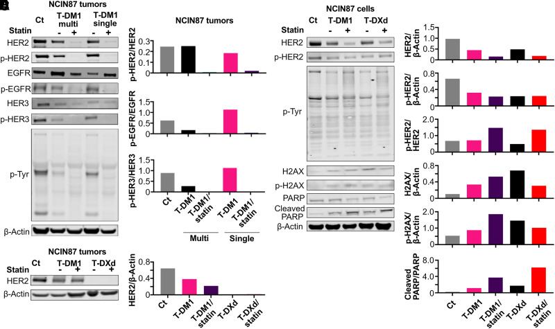
FIGURE 5.
(A and B) Western blot analyses (A) and quantification (B) at 43 d after treatment of NCIN87 tumors for control, T-DM1 multiple-treatment regime, T-DM1/statin multiple-treatment regime, T-DM1 single-treatment regime, and T-DM1/statin single-treatment regime. Western blots show expression of proteins in HER2 downstream signaling pathways including HER2, phosphor-HER2, EGFR, phosphor-EGFR, HER3, phosphor-HER3, and phosphor-tyrosine. β-actin was used as loading control. Quantifications shown in B relate to A, and analyses for additional repeats are shown in supplemental materials. (C) HER2 Western blot analyses and quantification after treatment of NCIN87 tumors for control, T-DM1 single-treatment regime, T-DM1/statin single-treatment regime, T-DXd single-treatment regime, and T-DXd/statin single-treatment regime. β-actin was used as loading control. (D) Western blot analyses and (E) quantification for NCIN87 cells treated with vehicle, T-DM1, T-DM1/statin, T-DXd, and T-DXd/statin for 48 h. Western blots show expression of HER2, phosphor-HER2, phosphor-tyrosine, H2AX, phosphor-H2AX, cleaved PARP, and PARP. β-actin was used as loading control. Full membranes of all repeats are shown in supplemental materials. p-Tyr = phosphor-tyrosine.

