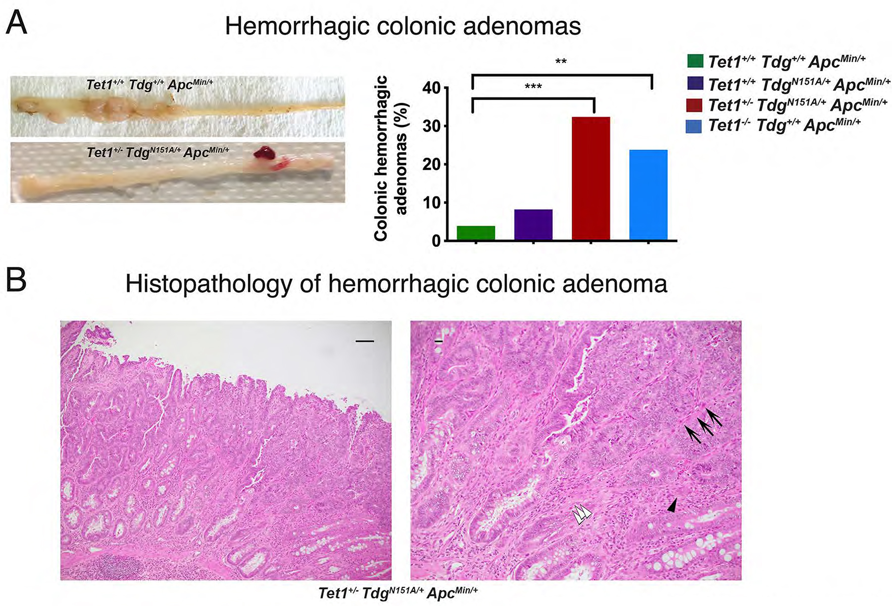
Figure 2 -. Hemorrhagic colonic adenomas in Tet1+/−TdgN151A/+ApcMin/+ mice and Tet1−/−Tdg+/+ApcMin/+ mice.

(A) Gross morphology and percent of hemorrhagic colonic adenomas in the four genotype-groups. **p-value=0.0044, ***p-value=0.0008. (B) Representative pictures of hematoxylin-eosin-stained sections of a hemorrhagic colonic adenoma developed in Tet1+/−TdgN151A/+ApcMin/+ mouse at 10x (left; scale bar=50 microns) and 20x magnification (right; scale bar=10 microns), showing evidence of malignant transformation, evidenced by overall disorganized architecture, irregular, ragged and invading glands. Frame in left panel corresponds to enlarged right panel. White arrowheads mark cancer cells invading the stroma; black arrowhead points to a budding gland; arrows point to capillaries responsible for the characteristic hemorrhagic macroscopic appearance.
