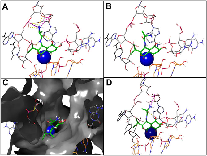
Fig. 2.
A Bactobolin A binds in a cleft between the 23S rRNA (gray sticks) and the tRNA (orange sticks), coordinating with a magnesium ion (blue sphere) and forming several internal and external hydrogen bonds (yellow dashes) and salt bridge interactions (magenta dashes). B The Bactobolin epimer is oriented away from the sub-pocket, interacting with different regions of the rRNA. C The space fill representation with the Bactobolin epimer suggests the potential to interrogate other sub-pockets. D The C4/C6 diastereomer regains interactions that were lost in the C4 epimer

