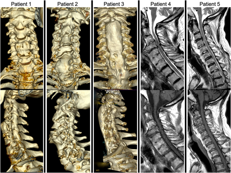
FIGURE 2.

Phenotypic variability. Panel of 5 patients from our cohort highlighting the heterogeneity of cervical DISH. 3-dimensional reconstructed computed tomography anterior-posterior and lateral views illustrate variability in disease severity. Patient 1 demonstrates mild DISH with osteophytes bridging from C4 to C6. Patient 2 has a discontinuous DISH pattern through the cervical spine with breaking at C5-6 resulting in esophageal compression and dysphagia. Patient 3 shows a more laminar, continuous pattern. T1-weighted and T2-weighted MRI images demonstrate variability in T2 hyperintensity of the ectopic bone mass. Patient 4 with DISH that is homogenous in intensity to native bone, whereas patient 5 has a remarkable amount of T2 hyperintensity in the ectopic bone mass in comparison with patient 4 and the patient's native bone. DISH, diffuse idiopathic skeletal hyperostosis.
