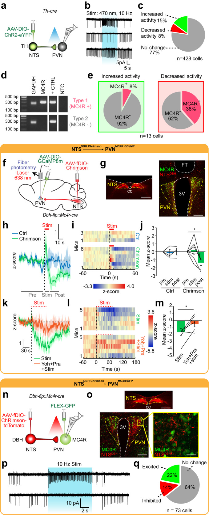
Fig. 5. Activating NTSTH → PVN connection produces heterogenous response profile in target neurons.

a Schematic for ChR2-assisted functional mapping of NTSTH → PVN connection using ex vivo electrophysiology. b Representative loose seal traces from PVN neurons during NTSTH axon photostimulation (blue shaded region). c Pie chart summarizing percent of PVN neuron response types to NTSTH photoactivation (n = 428 neurons, 6 mice). d Representative agarose gel of RT-PCR outcomes assessing MC4R expression from single PVN neurons collected following patch clamp recordings. e Summary of PVN MC4R+ and MC4R- neurons activated (n = 13 cells, 3 mice) or inhibited (n = 13 cells, 3 mice) by NTSTH axon photostimulation. f Schematic for assessing NTSDBH → PVNMC4R connection in vivo. g Photomicrograph of Chrimson (red) expression in NTSDBH soma and PVN projections, GCaMP8m (green) transduced PVNMC4R neurons, and fiber tip (FT) (scale bars: 500 μm, 200 μm). h–j Effect of NTSDBH → PVN projection photostimulation (10 s, 10 Hz) on PVNMC4R GCaMP8m fluorescence in control (n = 3 mice) or Chrimson transduced (n = 6 mice) animals. h Average traces, i individual temporal heatmaps, and j z-score quantification during pre-stimulation, stimulation (red line), and post-stimulation periods (2-way RM-ANOVA with Tukey’s multiple comparison, *p = 0.0141). k–m PVNMC4R GCaMP8m fluorescence during train photostimulation of NTSDBH → PVN axons before and after intraperitoneal yohimbine + prazosin cocktail administration (n = 5 mice). k Average traces, l individual temporal heatmaps, and m quantification during stimulation period (two-tailed, paired t-test, *p = 0.038). n Schematic for ex vivo cell-type specific Chrimson-assisted circuit mapping of NTSDBH → PVNMC4R connection. o Representative images showing expression of Chrimson in NTSDBH soma and PVN axons, and GFP transduced PVNMC4R (scale bars: 500 μm, 200 μm, 50 μm). p Representative loose seal traces and q summary of PVNMC4R neurons activated or inhibited by NTSDBH axon photostimulation (n = 73 neurons). Data are presented as mean values +/− SEM.
