Figure 3.
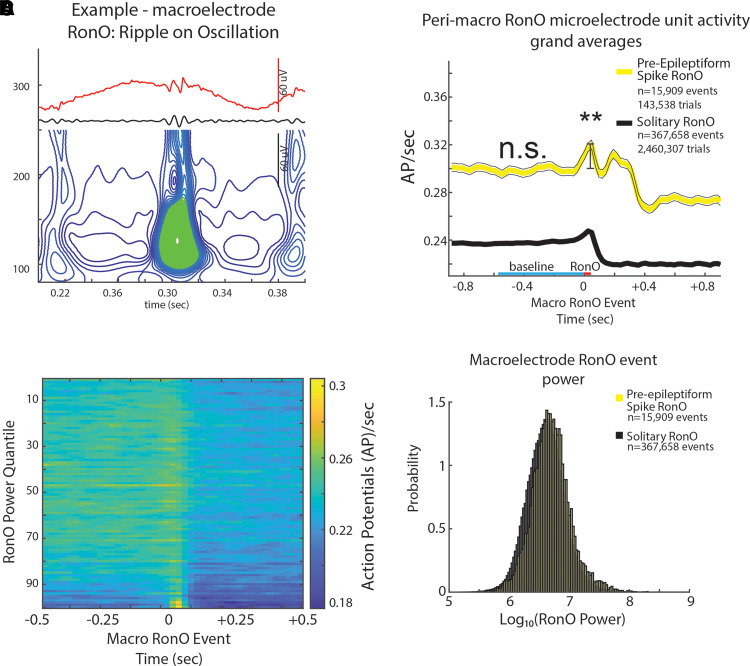
iEEG RonOs associated increases in unit firing rate are larger if the RonO precedes (<300 ms) an epileptiform spike. (A) An example RonO identified using the topographical analysis of the wavelet convolution. (B) Peri-RonO unit Gaussian smoothed AP firing rates from 174 units, which exhibited a significant RonO triggered increase in firing (P < 0.001, FDR corrected), averaged by quantiles defined by the log10(power) of the RonO events. Higher power RonOs are associated with a greater increase in AP firing rate (GLMM, , Supplementary Table 7). (C) Grand average of the Gaussian smoothed HFO event-unit AP train trials for all peri-RonO event trials not preceding an epileptiform discharge (black, dotted lines = 95% confidence interval), and peri-RonO event trials that did precede (<300 ms) an epileptiform discharge (yellow). Although the baseline (cyan line) firing rate appears different between the two conditions, no increase was detected in the GLMM that accounted for RonO power as a fixed effect and the random effects of patient, macroelectrode and unit [not significant (n.s.), P > 0.05, Supplementary Table 9]. RonO events that preceded an epileptiform discharge, relative to those that did not, had an increased peak AP firing rate during the RonO event (red line) compared with baseline (cyan line) (**, GLMM, P < 1e−10, Supplementary Table 6). (D) Normalized histogram of fRonO event power in macroelectrode recordings. RonO events preceding spikes had a larger power (t-test, P < 1e−10, Cohen’s d = 0.3) than solitary RonO, and the interaction of RonO event power and pre-spike status significantly predicted increased firing rate (GLMM, P < 0.01, Supplementary Table 7).

