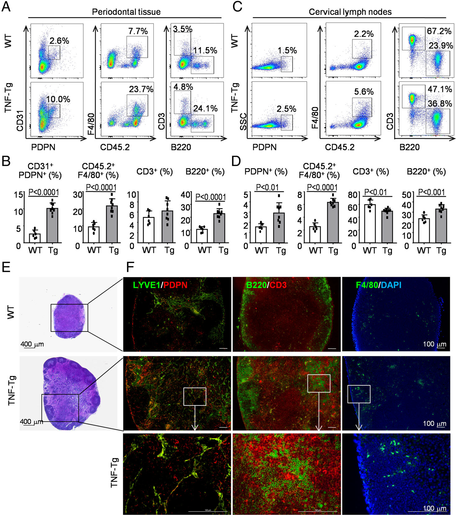
Figure 2.

Numbers of lymphatic endothelial cells, macrophages, and B cells are increased in periodontal tissues and in the cervical (draining) lymph nodes in RA-associated periodontitis. Six-month-old TNF-Tg RA mice and their WT littermates were used. (A) Cells from periodontal tissue were collected and analyzed by flow cytometry. Representative dot-plot shows the population of various cell subsets. (B) Subpopulations of cells in the periodontal tissue. (C) Cells from cervical lymph nodes were collected and analyzed by flow cytometry. Representative dot-plot shows the population of various cell subsets. (D) Subpopulations of cells in the cervical lymph nodes. Paraffin sections of cervical lymph nodes were H&E-stained (E) and adjacent sections were subjected to IF (F) for LYVE1 (green) and PDPN (red) for LVs (left-hand panel), for B220 (green) for B cells and CD3 (red) Ab for T cells (middle panel), with F4/80 (green) for macrophages (right-hand panel). Between six and eight mice per group were included in each experiment. Two-tailed unpaired Student’s t-tests were performed.
