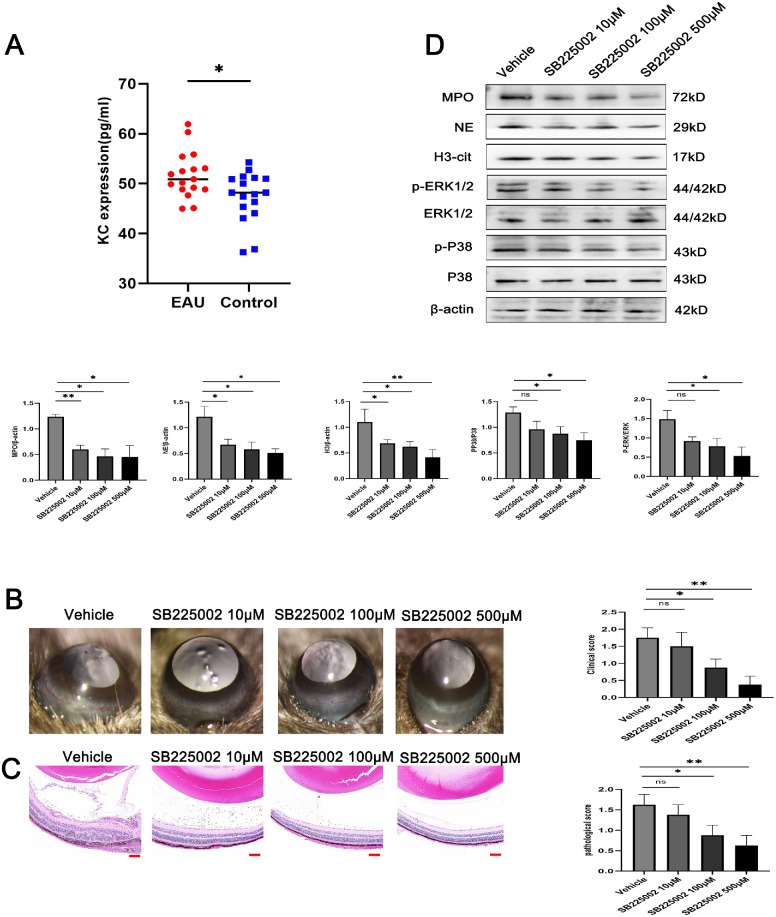
Figure 7.
SB225002 treatment ameliorated the clinical and histological manifestations in EAU mice. (A) Comparison of serum KC level between the EAU mice and control mice (n = 17 per group; unpaired t test). (B) Representative images of the slit lamp (left) and quantification of clinical scores (right) of the EAU mice treated with the CXCR2 inhibitor SB225002 (n = 4 per group; Kruskal–Wallis test). (C) Representative images of hematoxylin and eosin staining (left) and quantification of histopathological scores (right) of the EAU mice treated with the CXCR2 inhibitor SB225002 (n = 4 per group; Kruskal–Wallis test; scale bars, 25 µm). (D) Western blot for H3Cit, NE, MPO, ERK, p-ERK, P38, and p-P38 expression in retinas (n = 3 per group; one-way ANOVA test). The quantification of protein samples was determined via densitometry and normalized. The data were expressed as mean ± SD. *P < 0.05, **P < 0.01.

