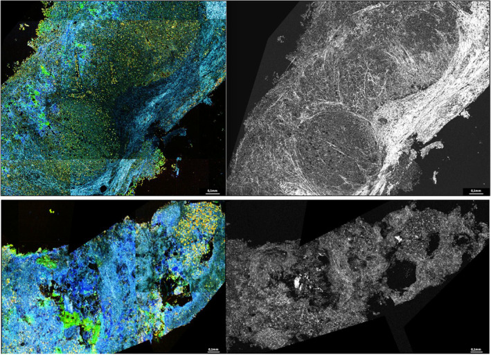
FIGURE A1.

Benign and malignant lymph nodes. In DCI, cells appear as yellow formation with black or red core, extracellular matrix appear as blue linear formations. We can notice a well‐defined lymphatic lobule on this benign lymph node image, the cortex is respected and no irregularity is noted. The cellular organization is regular within the lobule without pathological clusters. In contrast, the second specimen shows a complete disorganization with many irregularities. Clusters of pathological cells can be seen, particularly in the upper right‐hand corner of the image.
