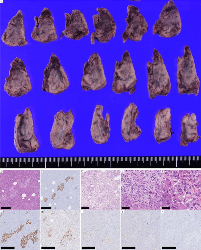
Fig. 1.

Pathological features of ANHH. (A) Macroscopic and (B) microscopic examinations (H&E) and (C) insulin immunostaining reveal no mass lesion in the sections of the resected pancreas. (D) Ductuloinsular complex. H&E. (E) Multiple β-cells with an enlarged and hyperchromatic nucleus and abundant clear cytoplasm. H&E. (F) Macronucleoli in β-cells. H&E. (G-J) Immunostaining. Islets with normal spatial distribution of the various cell types. (G) Insulin, (H) Glucagon, (I) Somatostatin, and (J) Pancreatic polypeptide staining. (K) Ki-67 immunostaining reveals no proliferative activity of the islet cells. Scale bars in B, C, G-K are 250 μm; the scale bar in D is 100 μm; the scale bar in E is 50 μm; and the scale bar in F is 25 μm
