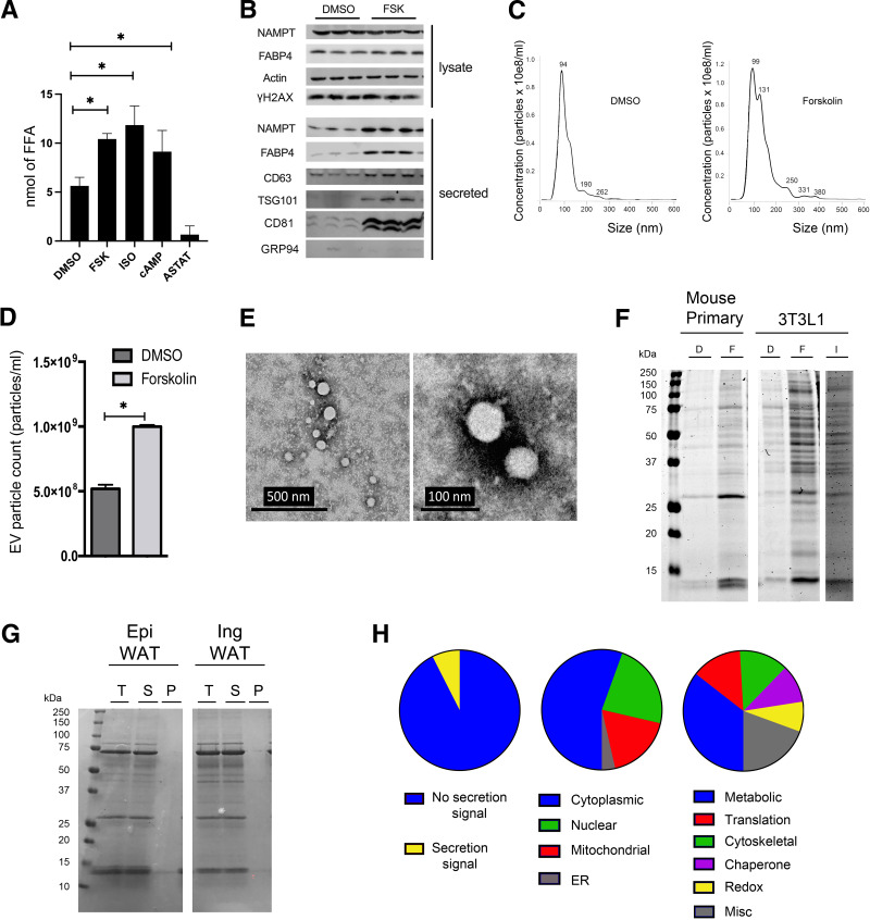
Figure 1.
FSK stimulates protein secretion. A: Differentiated 3T3-L1 adipocytes were treated with DMSO, 10 μmol/L FSK, 10 μmol/L Iso, 1 mmol/L 8-Br-cAMP (cAMP in the figure) or 15 μmol/L ATGL inhibitor (ASTAT) for 4 h. Free fatty acid (FFA) levels in cell culture medium were measured and quantified by colorimetric assay. B: Differentiated 3T3-L1 adipocytes were treated with DMSO or 20 μmol/L FSK for 4 h. Whole-cell lysates were harvested, and equal amounts of intracellular protein underwent SDS-PAGE for Western blot analyses. For the secreted proteins, equal volumes of cell culture medium were loaded for Western blot analyses (NAMPT and FABP4). For the AdEV markers CD63, TSG101, and CD81, AdEVs were pelleted from equal volumes of cell culture medium, using the Total AdEV isolation reagent (catalog no. 4478359, Invitrogen). Subsequently, the pellets were lysed and resuspended into equal volume of RIPA buffer before analyses via Western blots. C and D: Differentiated 3T3-L1 adipocytes were treated with DMSO or FSK for 4 h and Nanosight analyses (see experiment details in Research Design and Methods) was used to quantify particle numbers (C) and AdEV particle size (D). E: 3T3-L1 adipocytes were treated with FSK for 4 h and AdEVs were pelleted using the Total AdEV isolation reagent. The AdEVs were negatively stained and mounted on a grid for transmission electron microscopy (46). F: Mouse primary and 3T3-L1 adipocytes were treated with FSK or Iso for 2 h and equivalent volumes were analyzed on SDS-PAGE and stained with Coomassie protein stain. G: C57Bl/6J mice were sacrificed and WAT was minced and washed. The explants were incubated in Krebs Ringer Bicarbonate Hepes (KRH) buffer with 10 μmol/L FSK for 2 h. The medium was recovered and ultracentrifuged at 100,000g for 1 h. The pellet was resuspended in the same volume of KRH. Equivalent volumes total protein (T), soluble (S), and pellet (P) were separated by SDS-PAGE and stained with Coomassie stain. Epi, epididymal; Ing, inguinal. H: Analyses of the mass spectrometry results of total FSK-dependent protein secretion, including percentage with classic secretion signals, subcellular locations, and functional pathways. Results represent the mean ± SEM. *P < 0.05. EV, extracellular vesicle.

