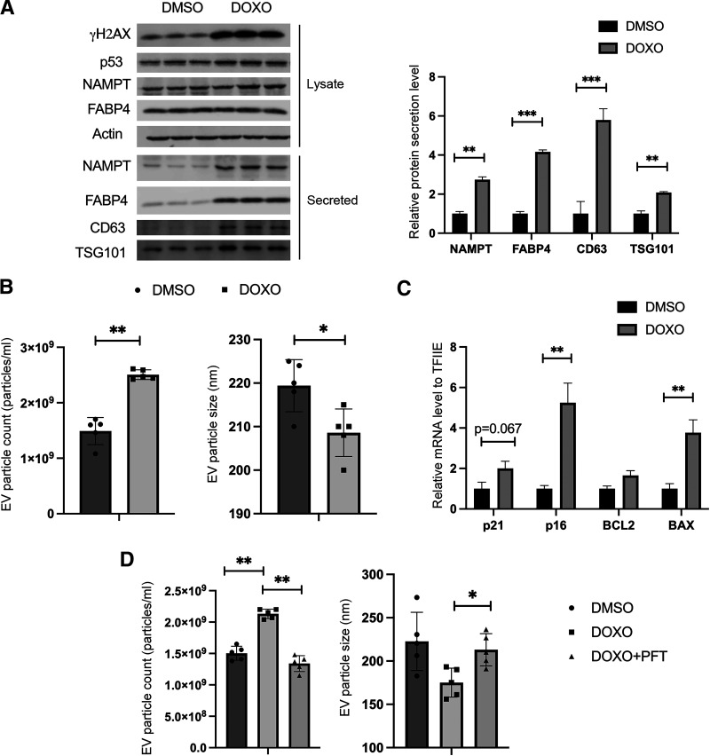
Figure 5.
DNA damage facilitates FABP4 and AdEV protein secretion from adipocytes in a p53-dependent manner. A: Differentiated 3T3-L1 adipocytes were treated with DMSO or 10 μmol/L DOXO for 24 h. Western blot analysis was used to measure intracellular levels of the proteins γH2AX, p53, NAMPT, FABP4, and actin, as well as secreted protein level (NAMPT, FABP4, CD63, and TSG101) in the cell culture medium. For the intracellular lysate, whole-cell lysates were harvested for each condition and equal amounts of intracellular protein were loaded to Western blots. For the secreted samples, equal volumes of cell culture medium were loaded onto Western blots. For the AdEV markers CD63 and TSG101, AdEVs were pelleted from an equal volume of cell culture medium, using the Total AdEV isolation reagent. The pellets were lysed and resuspended into an equal volume of RIPA buffer before loading to Western blots. B: Differentiated 3T3-L1 adipocytes were treated with DMSO or 10 μmol/L DOXO for 24 h. AdEVs were pelleted from an equal volume of cell culture medium and resuspended in 1 mL PBS for future analyses. AdEV size and amounts were measured by Nanosight analyses (see experiment details in Research Design and Methods). C: Differentiated 3T3-L1 adipocytes were treated with DMSO or 10 μmol/L DOXO for 24 h. The RNA was purified and p53 transcriptional targets expression was measured by qRT-PCR. D: Differentiated OP9 adipocytes were pretreated with DMSO or 20 μmol/L PFT for 2 h, then treated with DMSO or 10 μmol/L DOXO for 24 h. AdEVs were pelleted, purified from equal volume of cell culture medium from different treatment, and resuspended in 1 mL PBS for analysis. AdEV size and amounts were measured by Nanosight analysis (see experiment details in Research Design and Methods). All results represent the mean ± SEM. *P < 0.05, **P < 0.01, ***P < 0.001. EV, extracellular vesicle.

