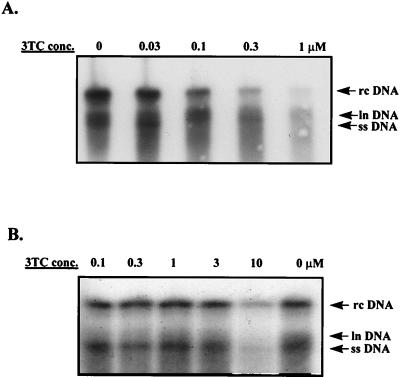
FIG. 2.
Sensitivity of the M539V variant to 3TC. HepG2 cells were transfected with pCMVhbv or pAT0304 and pZeoSV-Lacz. Twenty-four hours after transfection, the cells were treated with various concentrations of 3TC for 4 days. Intracellular viral core particles were isolated as previously described (6). Briefly, viral DNA was released from the core particles by proteinase K treatment followed by phenol extraction and ethanol precipitation. It was then separated by size by electrophoresis, transferred to a nylon filter, and detected with a radioactively labeled HBV-specific probe. The autoradiography exposure time was 16 h. (A) Core-associated HBV DNA isolated from HepG2 cells transfected with pCMVhbv and treated with various concentrations of 3TC. (B) Core-associated HBV DNA isolated from HepG2 cells transfected with pAT0304 and treated with various concentrations of 3TC. rc, relaxed circular; ln, linear; ss, single stranded. Note that there was a 3.5-fold difference in the amount of HBV DNA in the 0 drug lane in panels A and B after normalization to transfection efficiency.

