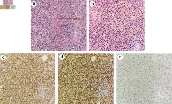
Figure 2.
Pathological images of the tumor. (a) (H&E, × 200) and (b) (H&E, × 400) show the morphology of tumor cells. Immunohistochemical findings. The tumor cells expressed CD38 (c, original magnification × 200) and kappa (d, original magnification × 200) without expression of lambda (e, original magnification × 200). H&E: hematoxylin and eosin.

