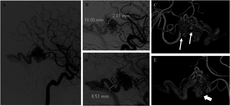
Figure 1.
(A) bAVM with a compact nidus receiving arterial supply mainly by terminal feeders arising from the right middle cerebral artery. (B) Demonstration of the measurement of the diameter of the major arterial feeder at 1 cm from the nidus. (C) Multiple intranidal aneurysms (arrows). (D) Single ectasic venous collector draining into the right transverse sinus (superficial venous drainage). (E) 3D DSA reconstruction demonstrating the presence of a venous varix (arrow) at the origin of the venous collector.

