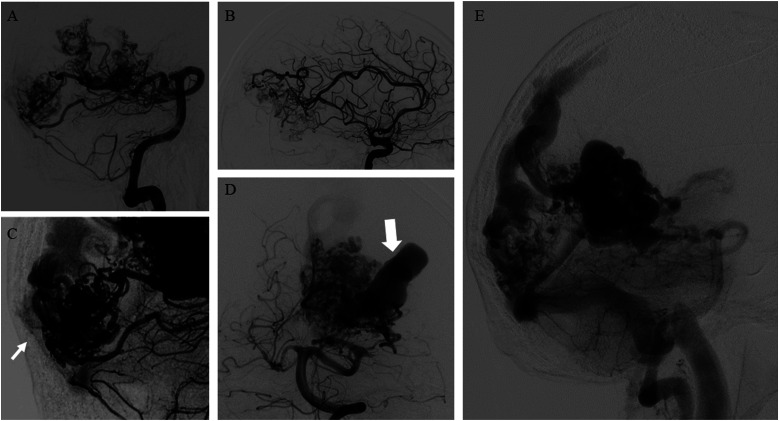
Figure 3.
(A–B) bAVM with a plurifocal and diffuse nidus, receiving arterial supply by both anterior and posterior circulations. (C) Pials arterial feeders (arrow) also supply the plurifocal nidus of bAVM. (D) Presence of an intranidal giant aneurysm (arrow). (E) Mixed (superficial and deep) venous drainage draining the bAVM through cortical venous system and deep veins (the internal cerebral veins and the basal veins).

