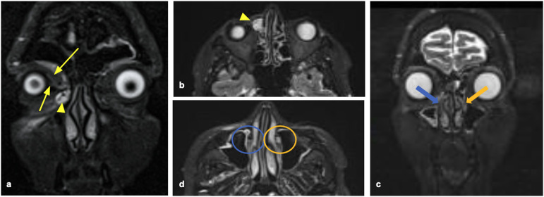
Figure 2.
65-year-old female patient affected by right recurrent dacryocystitis. The T2-weighted image (a) showed a dilated lacrimal sac (arrowhead), with mild dilation of the inferior and superior canaliculi (white arrows). Axial (b, d) and coronal (c) reconstructions of the STIR images. In (b) the dilatation of the lacrimal sac is detectable (arrowhead). The left nasolacrimal duct is regularly filled with fluid (c, orange arrow), whereas the right nasolacrimal duct is empty (blue arrow): this finding is also visible in (d), where the axial image shows the fluid content of the left nasolacrimal duct (orange circle), whereas the left one is empty (blue circle). The obstruction was diagnosed in the nasolacrimal duct and confirmed by the endoscopy.

