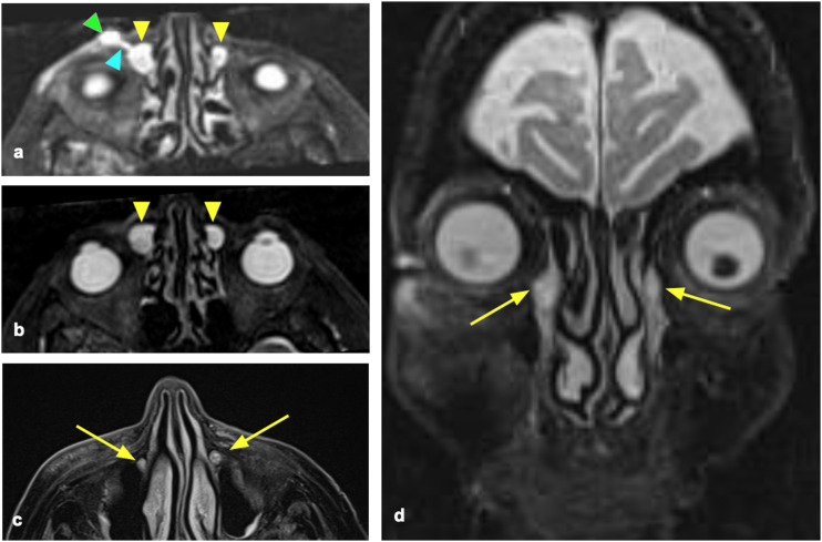
Figure 3.
73-year-old female patient affected by bilateral recurrent dacryocystitis. Axial (a, b, c) and coronal (d) reconstructions of the STIR images. Bilateral dilatation of the lacrimal sacs (a, b) (yellow arrowheads). The right lacrimal ducts present a fistulization (light blue arrowhead) with subcutaneous fluid collection (green arrowhead) (a). Both nasolacrimal ducts are filled with fluid (c, d, yellow arrows). The stenosis was diagnosed as located in the lacrimal sac and confirmed by endoscopy.

