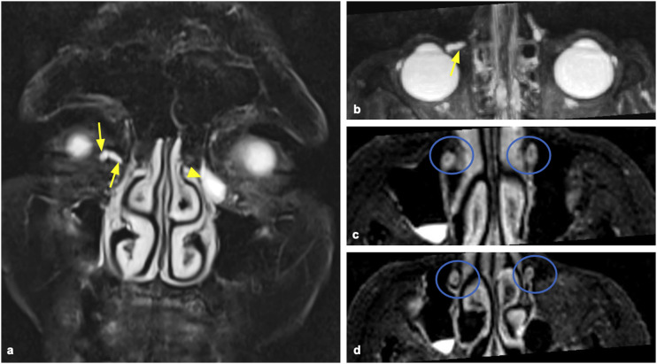
Figure 4.
Coronal MIP reconstruction of the heavily T2-weighted 3D coronal sequence (a) and axial MIP reconstruction of the heavily T2-weighted 3D coronal sequence (b) show stenosis of the common right canaliculus, with retro dilation of the common and inferior canaliculi (yellow arrows). Contralateral dilatation of the lacrimal sac (yellow arrowhead). Axial reconstructions of the 3D STIR sequence (c, d) show bilateral empty lacrimal ducts (blue circles). The patient was diagnosed as having stenosis located at the right common canaliculus and stenosis located at the left lacrimal sac.

