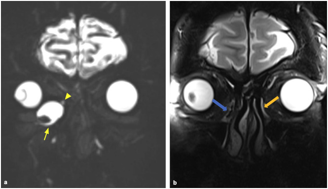
Figure 5.
A 42-year-old patient with recurrent episodes of right dacryocystitis. (a) Huge distension of the right lacrimal sac (yellow arrow), with evidence of stenosis (yellow arrowhead) at the level with the conjunction to the nasolacrimal duct. (b) Shows that the right duct is empty (blue arrow), whereas the left one is filled with fluid (orange arrow). In Supplementary Materials, we uploaded the video of the surgical procedure.

