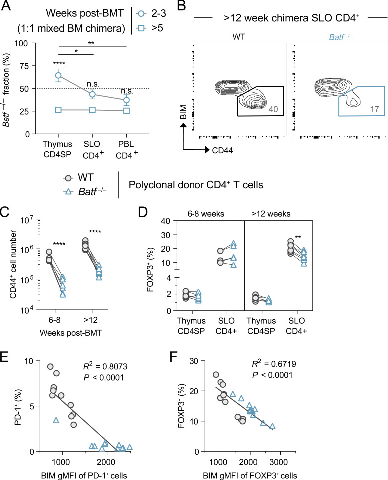
Figure S4.
Expression of BIM, CD44, FOXP3, and PD-1 is dysregulated in the Batf−/− donor-derived polyclonal CD4+ T cells observed in 1:1 WT and Batf−/− mixed BM chimeric mice. (A) Kinetics of relative T cell reconstitution in thymus, SLOs, and blood of mixed BM chimeric mice. Data represent the mean ± SD percentage of Batf−/− donor-derived T cells in samples, with significant differences between time points or tissue source as indicated (one-way ANOVA; *P < 0.05, **P < 0.01, ****P < 0.0001, n.s. = not significant). (B) Representative CD44+ BIMlo polyclonal CD4 T cell SLO accumulation in week 12 mixed BM chimeras. (C and D) Quantitation of CD44+ conventional (C) and FOXP3+ Treg (D) cell generation over time. Lines connect donor cell groups of individual mixed BM chimeric mice, with significant differences between WT (circles) and Batf−/− (triangles) as indicated (paired t test; **P < 0.01, ****P < 0.0001). (E and F) Correlation observed between BIM expression and the frequency of PD-1+ conventional (E) or FOXP3+ Treg (F) cells in both WT (circles) and Batf−/− (triangles) donor-derived populations. Source data as in Fig. 8, A and B.

