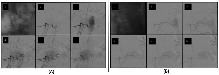
Figure 8. Spinal angiogram from case three.
Sequential images from the spinal angiogram of a right L1 radicular branch. The nidus of the dural AVF is fed from a linear ascending artery off the right L1 radicular branch and drains into veins on the posterior aspect of the spinal cord. (A) Anterior/posterior view; (B) Lateral view.

