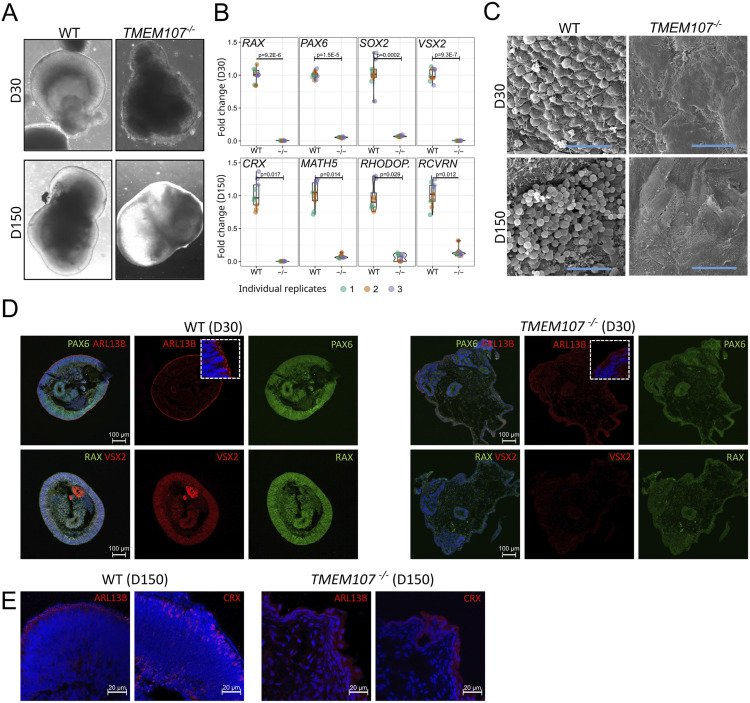
Figure 4. TMEM107 is essential for human retinal development.
(A) Morphology of retinal organoids at D30 and D150, as demonstrated using brightfield microscopy. (B) Expression of retinal genes at early stage (D30—upper row) and late stage (D150—bottom row) of the differentiation process in WT and TMEM107−/− retinal organoids, as demonstrated using RT–qPCR; parametric paired, two-tailed t test; n = 3. (C) Microphotograph of retinal organoid surface (D30—upper row, D150—bottom row), as demonstrated using SEM. Scale bars represent 10 μm. (D) Expression of PAX6 (green), ARL13B (red), RAX (green), and VSX2 (red) in WT and TMEM107−/− retinal organoids at D30, as demonstrated using immunofluorescence staining. Nuclei are counterstained with DAPI. (E) Expression of ARL13B and CRX in WT and TMEM107−/− retinal organoids, as demonstrated using immunofluorescence staining. Nuclei are counterstained with DAPI.

