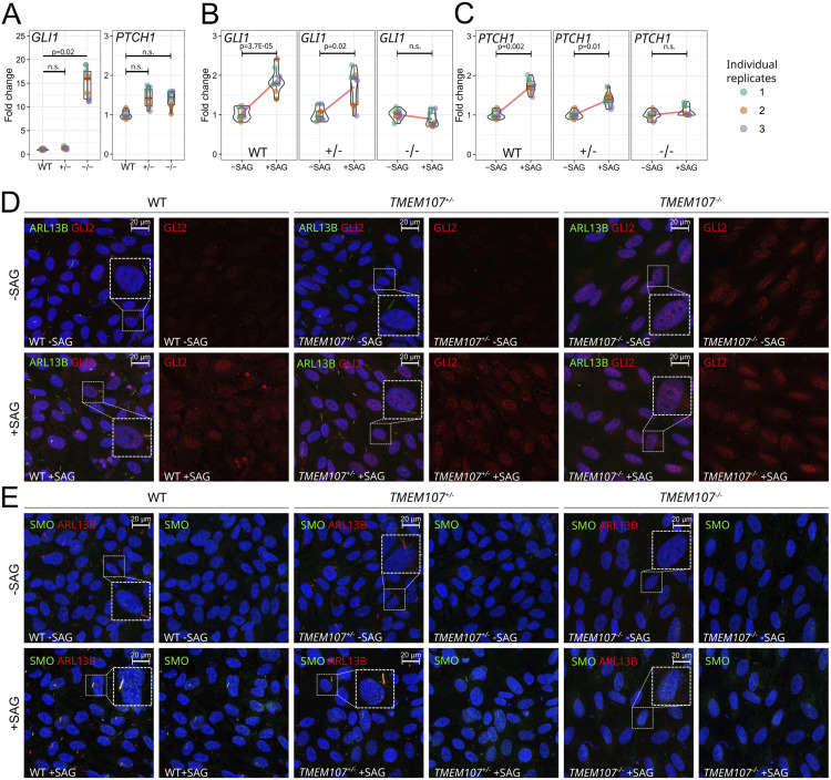
Figure 6. Loss of TMEM107 leads to aberrant Shh signaling in retinal cells.
(A) Expression of GLI1 and PTCH1 in WT, TMEM107+/−, and TMEM107−/− ARPE-19 cells, as demonstrated using RT–qPCR; Paired, two-tailed t test; n.s. = non-significant; n = 3. (B, C) Expression of GLI1 and PTCH1 upon SAG treatment in WT, TMEM107+/−, and TMEM107−/− ARPE-19 cells, as demonstrated using RT–qPCR; Paired, two-tailed t test; n.s., nonsignificant; n = 3. (D) Expression of ARL13B (green) and GLI2 (red) upon SAG treatment in WT, TMEM107−/−, and TMEM107+/− ARPE-19 cells, as demonstrated using immunofluorescence staining. (E) Expression of ARL13B (red) and SMO (green) upon SAG treatment in WT, TMEM107−/−, and TMEM107+/− ARPE-19 cells. Nuclei are counterstained with DAPI (blue).

