
Fig. 5. HIF-1α synchronizes YAP nuclear translocation in HIF-1α knockdown, knockout and overexpressed cells.
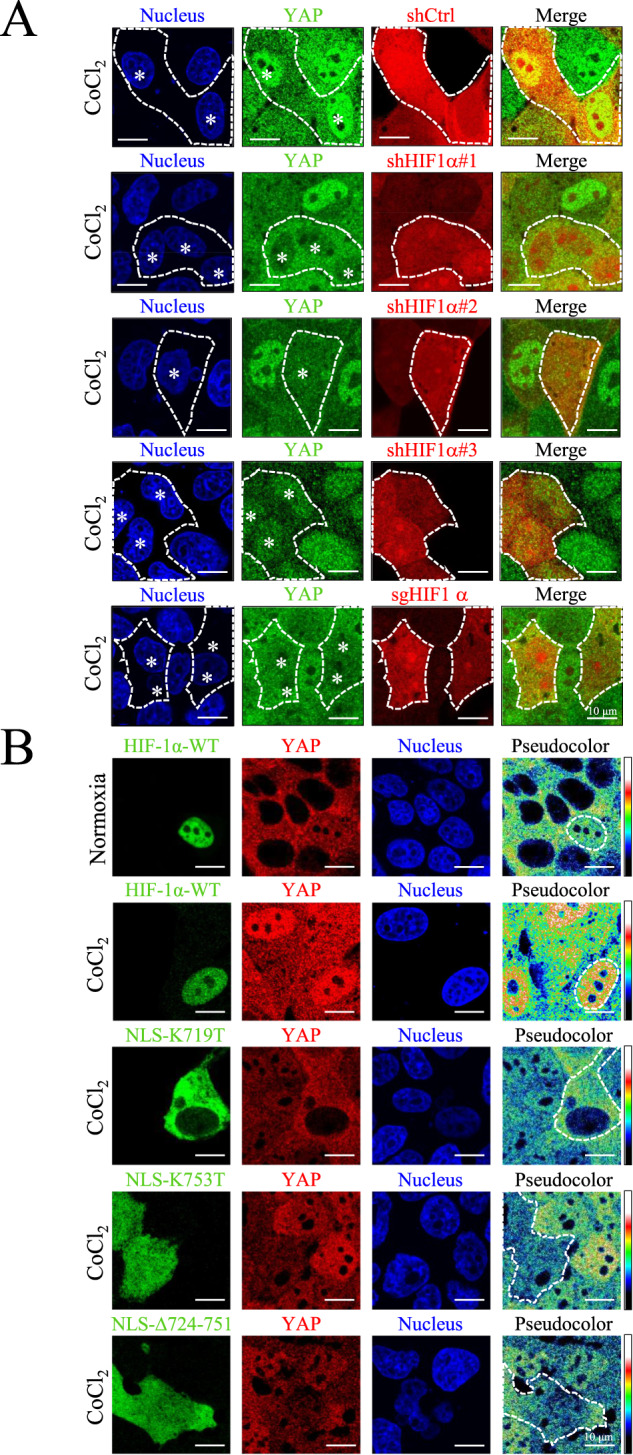
A High-density MDCK-shCtrl, MDCK-shHIF-1α #1, #2, #3, and MDCK-sgHIF-1α cells were treated with 400 μM CoCl2 for 8 h. Immunofluorescence images of YAP (green), shCtrl cells (red), shHIF-1α cells (red), and sgHIF-1α cells (red) were obtained using a confocal microscope. White dashed lines indicate the border of shCtrl, shHIF-1α and sgHIF-1α cells. White stars indicate the nuclei of shCtrl, shHIF-1α and sgHIF-1α cells. Scale bar: 10 μm. B Cells were either transient transfected with HIF-1α-WT-GFP (in MDCK-parental cells) or HIF-1α-NLS mutants: HIF-1α-NLS-K719T-GFP, HIF-1α-NLS-K753T-GFP, and HIF-1α-NLS-Δ724-751-GFP (in MDCK-shHIF-1α cells). The transfected MDCK-parental cells were then subjected to normoxia and 400 μM CoCl2, and the transfected MDCK-shHIF-1α cells were subjected to 400 μM CoCl2, respectively. After 8 h, images of HIF-1α (green), YAP (red and pseudo-color), and nuclei (blue) were captured using confocal microscope. Pseudocolor images show expression pattern of YAP. White dashed lines indicate the border of exogenous HIF-1α variants. Scale bar: 10 μm.