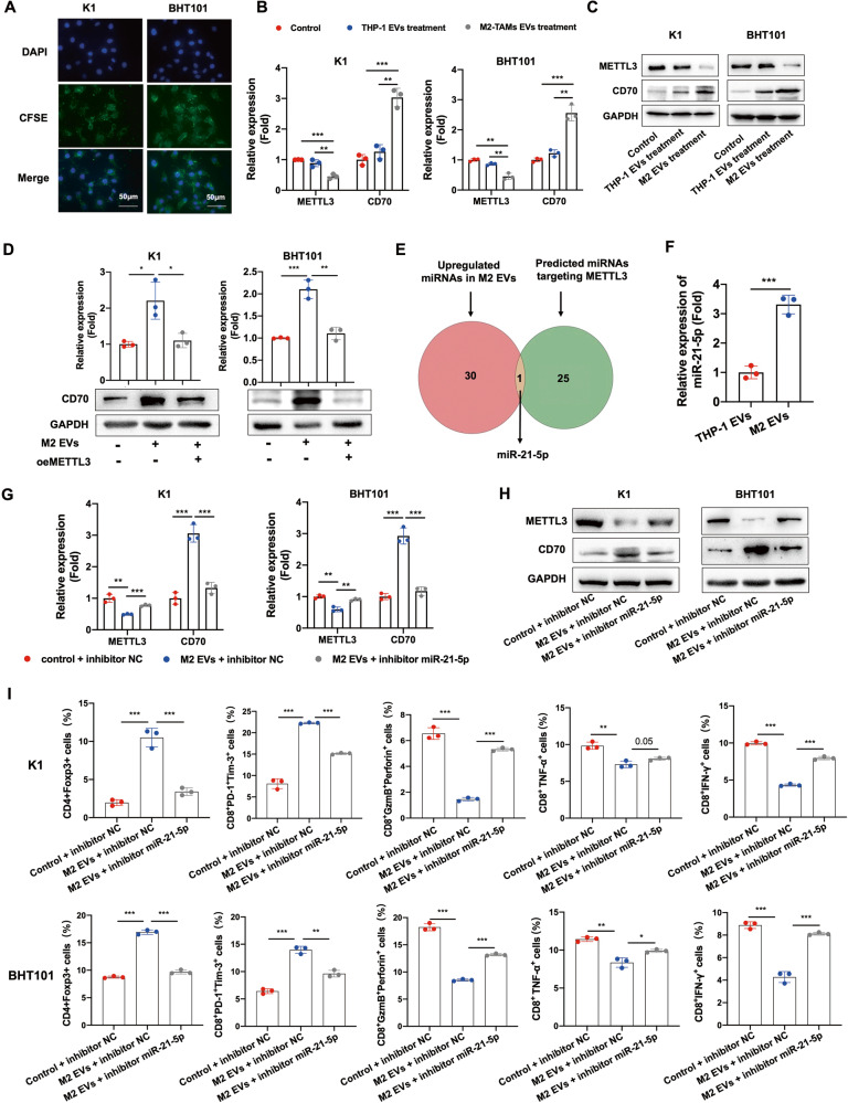
Fig. 5. M2 macrophage-derived EVs downregulated METTL3 in PTC and ATC cells through miR-21-5p.
A Representative images of K1 and BHT101 cells with phagocytosed CSFE-labeled M2 EVs (scale bars, 50 μm). The cell nucleus were stained with DAPI. B, C METTL3 and CD70 mRNA and protein levels in K1 and BHT101 cells co-cultured with M2 EVs, co-cultured with THP-1 cells and cultured alone. D CD70 mRNA and protein levels in K1oeMETTL3 and BHT101 oeMETTL3 cells co-cultured with M2 EVs. E Overlap between upregulated miRNAs in M2 EVs and predicted miRNAs targeting METTL3. F The expression of miR-21-5p in THP-1 EVs and M2 EVs. G, H METTL3 and CD70 mRNA and protein levels in K1 and BHT101 cells co-cultured with M2 EVs in the presence or absence of the miR-21-5p inhibitor. I Percentage of CD4+Foxp3+ Tregs, CD8+PD-1+Tim-3+ terminally exhausted T cells, CD8+GzmB+perforin+ T cells, CD8+TNF-α+ T cells and CD8+IFN-γ+ T cells in PBMCs co-cultured with M2 EVs-treated K1 and BHT101 cells expressing the miR-21-5p inhibitor. Data of are presented as the mean ± SD of three independent experiments. The p-values in (B), (D), (F), (G), (H) and (I) were calculated by Student’s t-test. *p < 0.05; **p < 0.01; ***p < 0.001.

