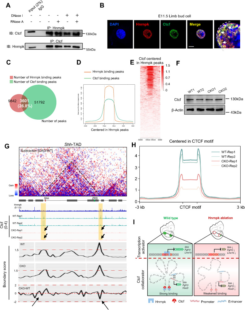
Fig. 6. Hnrnpk weakens the binding strength of Ctcf at TAD boundaries.
A Co-Immunoprecipitation (co-IP) assay between Hnrnpk and Ctcf at E11.5 in WT limb bud after treatment with PBS, DNase I, RNase A, or both. B Subcellular location of Hnrnpk and Ctcf at E11.5 in primary WT limb bud cell. The white arrows indicated the overlapped Hnrnpk and Ctcf signals in the nucleus. The white dotted line indicated the margin of nucleus. Scale bar: 10 μm. C The Hnrnpk- and Ctcf- binding peaks at E11.5 in WT limb buds and their overlap. The ratio indicated the proportion of overlapped peaks in the total peaks of Hnrnpk. D, E The binding profile of Ctcf centered in Hnrnpk binding peaks. F Protein level of Ctcf at E11.5 in WT and CKO limb buds. β-Actin was used as the loading control. G Hi-C contact map (upper) and genome browser tracks of Hnrnpk and Ctcf CUT&RUN-seq data (bottom) at Shh-TAD. The black arrows indicated the decreased binding strength of Ctcf at E11.5 in CKO limb bud. The yellow lines indicated the boundaries at both sides of Shh-TAD. H The Ctcf binding peaks centered in CTCF-motif at E11.5 in WT and CKO limb buds. I A proposed model illustrated the role of Hnrnpk as a transcription activator and insulator protein collaborator, which regulated the transcription of key regulatory genes during embryonic limb bud development.

