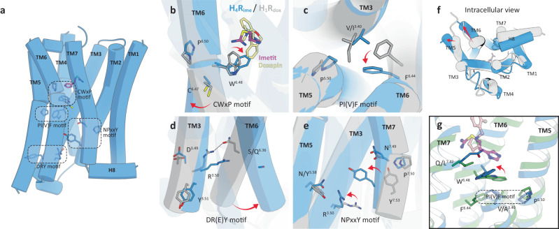
Fig. 5. Activation mechanism of H4R.
a Activation motifs in the H4R structure. Each motif residue is shown as a blue stick. b–g Structural comparison between the active H4Rime (blue) and antagonist-bound inactive H1Rhis (gray, PDB 3RZE). (b) CwxP motif, (c) PI(V)F motif, (d) DR(E)Y motif, (e) NPxxY motif. Imetit and doxepin are shown as magenta and khaki sticks. The residues involved in receptor activation are indicated by sticks, and conformational changes are shown as red arrows. Microswitch rearrangement allowed movement outside the cytoplasmic ends of TM5 and 6 (f). g Gln3477.42 coordination and its effect on surrounding residues.

