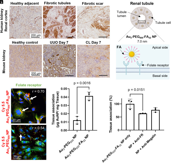
Fig. 2.
In vitro and ex vivo binding of Au3-PEG500-FA32 NPs to folate receptor (FR) on renal tubules after 2 h postincubation of cells or tissue blocks with NPs. (A) IHC staining of kidney biopsies from patients with renal fibrosis showed that some fibrotic tubules have higher expression of FR (brown) than adjacent healthy tissues. Representative images from two kidney sections from each tissue group (healthy adjacent vs. fibrotic tubules vs. fibrotic scar). (B) In mice, IHC staining showed that the elevated expression of FR (brown) in selected tubule cells in the UUO kidney is distinct from the diffuse, homogenous FR expression in healthy and CL kidneys. Representative images from three kidney sections from each mouse, n = 3 mice/group. (C) Schematic of Au3-PEG500-FA32 NP binding to FRs on the lumen side of a tubule cell. (D) Representative confocal images show stronger in vitro association of Cy5.5-labeled Au3-PEG500-FA32 NP (red; with white arrows) to FR (green) on healthy mouse primary renal tubule cells than Au3-PEG500 NP. r: Pearson’s colocalization coefficient. (E) Ex vivo association of Au3-PEG500 and Au3-PEG500-FA32 NPs to freshly diced UUO kidney blocks. Statistical significance was evaluated using the Mann–Whitney U test. Error bar denotes 1 SD, with n = 3 across one experiment. (F) Ex vivo competitive binding assay. Diced UUO kidney blocks were incubated with Au3-PEG500-FA32 NP with or without antibody against FR or megalin. Statistical significance was evaluated using one-way ANOVA with Tukey’s post hoc test for multiple comparison. All P values ≤ 0.05 are displayed on the graphs. All bars and error bars represent mean ± SD.

