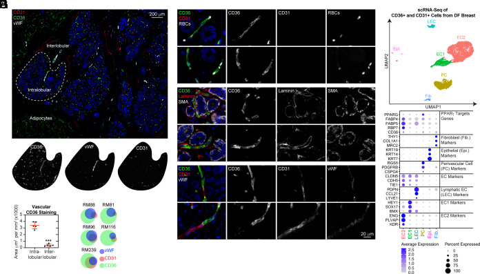
Fig. 1.
CD36 identifies intralobular capillaries lacking CD31 and vWF immunopositivity in the DF breast. (A) DF breast tissue from five donors aged 16–53 were subjected to mIHC analysis of CD36, CD31, and vWF. (B) The CD36-positive staining area, excluding adipocytes, was determined and normalized to the total area measured for the intralobular and interlobular stroma. (C) Following cell segmentation, vascular cells were scored and binned based on staining for each marker (CD36+, CD31+, and/or vWF+). Area-proportional Euler diagrams demonstrate the relationships between markers (overlaps between circles) and the proportion of each marker expressed within the intralobular vasculature (area of the circle). (D–F) DF breast was subjected to mIHC analysis for the additional markers validating vascular identity. (G) CD31+ and/or CD36+ from the DF breast (n = 14; median age: 24; range: 19–36) were subjected to MULTI-seq analysis. UMAP projection of cell clusters is shown. (H) A dot plot of selected marker genes indicative of cell cluster identity is shown. The size of the dot indicates the percentage of cells expressing the gene in each cluster, and the color of the dot represents the average expression level.

