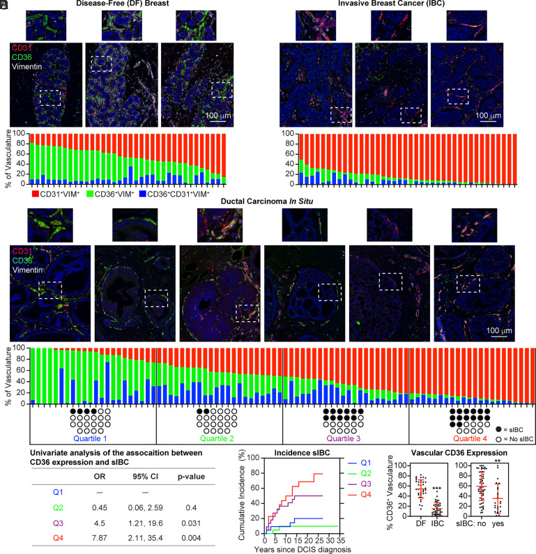
Fig. 3.
Loss of CD36 immunopositivity in the vasculature during breast cancer progression. (A) DF breast specimens (n = 36; median age: 30; range 16–78) were subjected to mIHC for CD36, CD31, and vimentin. The proportion of the total vasculature measured in the imaging area expressing CD36+ (green), CD31+ (red), or CD31+CD36+ (blue) is graphed. (B) 40 IBC specimens (median age: 52; range 30–79) were subjected to mIHC. (C) DCIS specimens (n = 88; median age: 57; range: 22–86) were subjected to mIHC. The patient population was split into quartiles based on the proportion of vasculature expressing CD36. Black circles indicate the development of subsequent IBC (sIBC) (median follow-up cases: 5 y, range: 1–16; controls: 19 y, range: 1–34). (D) Logistic regression was used to estimate associations between the proportion of breast vasculature expressing CD36 and sIBC. The odds ratio (OR) and 95% CI were calculated. (E) The cumulative incidence of sIBC following DCIS diagnosis is graphed for each quartile. (F) The total percentage of CD36-expressing vascular phenotypes (CD36+ and CD31+CD36+) in DF and IBC specimens, as well as DCIS cases (sIBC) and controls (no sIBC), is graphed.

