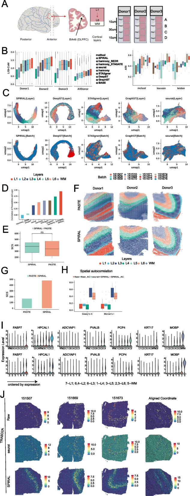
Fig. 3.

DLPFC. A Schematic of DLPFC data, which included four sequential sections A, B, C, and D of three donors. B The accuracy of batch effect removing and Louvain clustering, LISI-CoM (left) and ARI (right), in 13 integration experiments. C UMAP visualizations of 12-section integrations by different methods. Colors represent cell types (upper) and batches (bottom). D The barplot of Pearson correlation between deduced pseudo-time and annotated layers for 12-section integration experiment. E The boxplot of SCS of annotated layers on aligned coordinates by SPIRAL and PASTE in the 12 experiments. The “ns” means not significant differences. F Layers assignments on aligned coordinates by SPIRAL and PASTE for four sections of each donor. Colors represent different layers (upper) and shapes represent different sections. G SCS of annotated layers on aligned coordinates by SPIRAL and PASTE for 12-section integrations. H Spatial autocorrelations (Geary’s C and Moran’s I) of raw, SPIRAL, and Seurat-integrated gene expressions on original coordinates and raw and SPIRAL-integrated expressions on aligned coordinates, labeled as Raw, SPIRAL and Seurat, Raw-AC and SPIRAL-AC. I Violin plot of TRABD2A in annotated layers (upper) and deduced clusters (mclust, bottom). J spatial arrangements of raw (upper), Seurat (middle), and SPIRAL (bottom)-integrated gene expressions in original three spatial coordinates (the first three columns from the left) and aligned coordinates (the fourth column from the left)
