. 2023 Sep 21;15(9):e45696. doi: 10.7759/cureus.45696

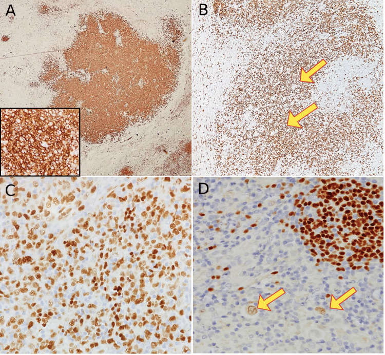
Figure 3. Immunohistochemistry studies done on the excisional lymph node diagnosed with composite classical Hodgkin lymphoma and diffuse large B-cell lymphoma. (A) CD20 immunostaining highlights strong expression in diffuse sheets of large, atypical cells; a component of diffuse large B-cell lymphoma (4x), high power magnification (40x) (inset); (B) CD20 in the adjacent area were negative in RS-cells (yellow arrow), note the positive background of small B-lymphocytes (4x); (C) PAX-5 shows strong nuclear staining in diffuse large B-cell lymphoma component (20x); (D) PAX-5 shows dim nuclear staining in RS cells confirming the classical Hodgkin lymphoma component (yellow arrow) (20x).

Figure 3