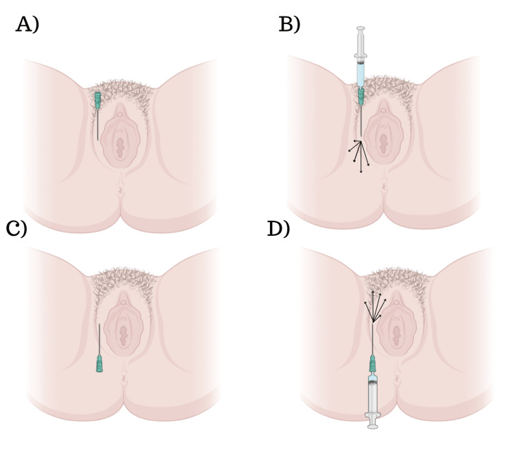
Figure 1. A schematic representation of our technique.
A) Creation of the orifice using a needle, followed by insertion of the microcannula.
B) Attachment of the syringe to the microcannula and retro-injection of the product. Note that the arrows indicate the different directions of product delivery, and it should be noted that the technique does not involve injecting the mucosa area.
C-D) Illustration of the same procedures applied to the upper part of the external vulva.

