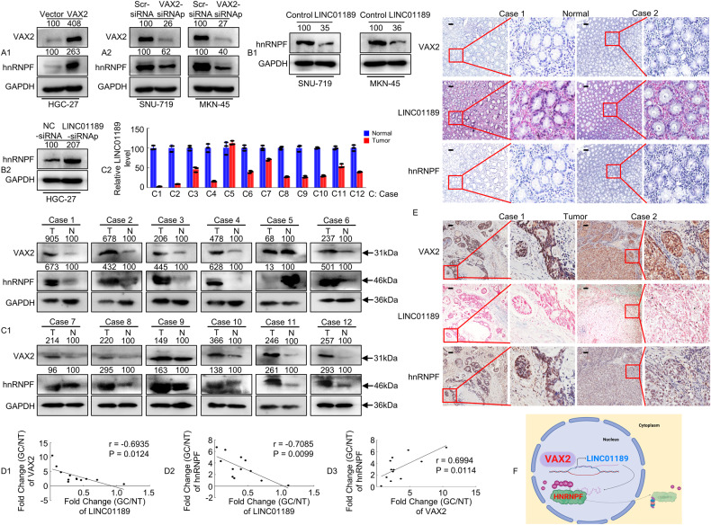
Fig. 7. Identification of a VAX2-LINC01189-hnRNPF axis in primary human GC samples.
A1/2, B1/2 VAX2 and/or hnRNPF expression detected using western blotting in GC cells transfected with Vector and VAX2 (A1), Scr-siRNA and VAX2-siRNAp (A2), Control and LINC01189 (B1), or NC-siRNA and LINC01189-siRNAp (B2). C1/2 The average tumor/normal epithelium (T/N) expression ratios of VAX2, hnRNPF, and LINC01189 detected using western blotting (C1) or qPCR (C2) in 12 paired GC tissues. C, case. D1-3 Correlation between VAX2 and LINC001189 expression levels, between LINC01189 and hnRNPF expression levels, or between VAX2 and hnRNPF expression levels in 12 GC tissues. Correlation coefficient (r) and P-value were acquired by Pearson correlation. E IHC and ISH were carried out to determine VAX2, LINC01189, and hnRNPF expression patterns in human GC tissues and normal epithelium tissues. Scale bar: 100 µm. F Proposed functional action of VAX2-LINC01189-hnRNPF axis in modulating GC progression.

