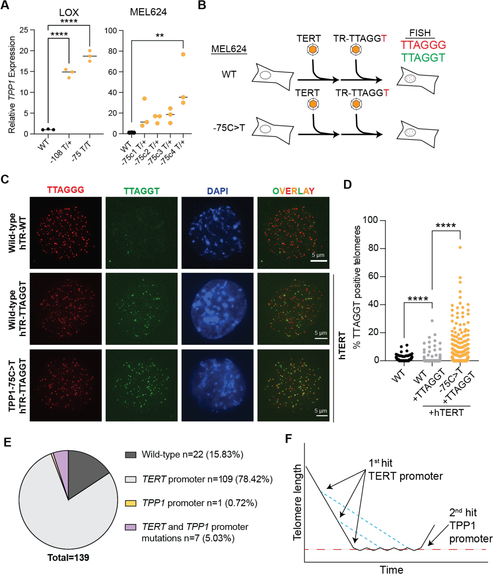
Fig. 4. TPP1 promoter mutations increase expression of the endogenous transcript and co-occur with TERT promoter mutations.

(A) Quantitative PCR of TPP1 expression following introduction of promoter mutations in LOX and MEL624 cells. Labels below the graph indicate the presumed zygosity based on sequencing. The media is shown from three independent measurements from each clone and groups were compared using one-way ANOVA followed by Dunnett’s multiple comparison test. (B) Schematic of the experimental approach to measure telomerase activity in genetically modified cells. Cells are transduced with a TERT-expressing lentivirus to increase the rate of variant telomere incorporation. Following introduction of the mutant telomerase RNA (encoding TTAGGT), cells are passaged and the canonical and variant telomeres are quantitated. (C) Fluorescent in situ hybridization for the WT (TTAGGG; red) and variant (TTAGGT; green) in parental or genome edited MEL624 cells. Images were taken 7 days after transduction with lentiviruses. (D) Quantitation of the fraction of telomeres that had both TTAGGG and TTAGGT signals from a single clone. Groups were compared using ANOVA with Dunnett’s correction for multiple comparison. **P<0.01 and ****P < 0.0001. (E) Proportion of cutaneous melanomas that had TERT, TPP1, or TERT+TPP1 variants from Hayward et. al. (25) (F) Model of telomere length dynamics in melanoma progression. TERT promoter variants likely occur early and slow telomere attrition but are not sufficient to prevent telomere shortening (blue dashed lines in model). Telomere shortening continues until cells enter crisis (red dashed line). Additional mutations, like the TPP1 promoter, are likely required to fully maintain telomeres and escape crisis (2nd hit).
