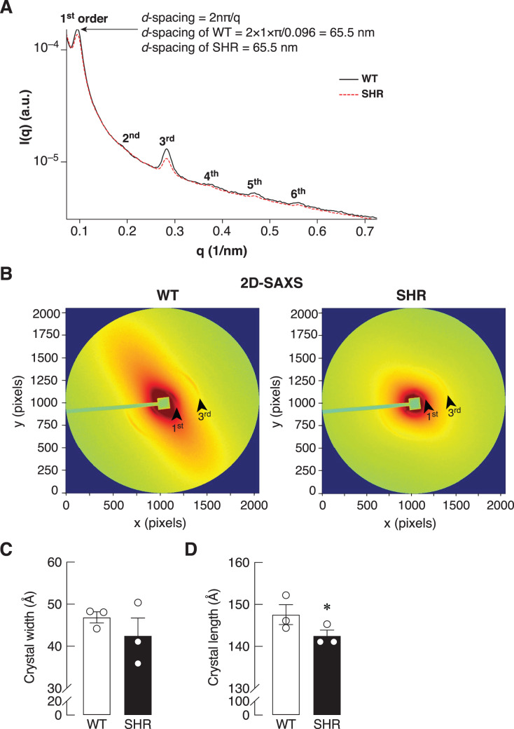
Figure 7. SAXS/WAXS indicated misalignment of bone collagen fiber in SHR.
Simultaneous small- and wide-angle X-ray scattering (synchrotron SAXS/WAXS) analyses of collagen fibril arrangement and crystallite dimensions in the cortical bone specimens from 24-week-old WT and SHR. (A) 1st–6th peaks from SAXS analysis. (B) 2D-SAXS analysis. Arrowheads represent 1st and 3rd peaks corresponding to those indicated in Fig. 7A. (C) WAXS analysis of crystal width, and (D) WAXS analysis of crystal length. The measurement of the crystal width and length are presented in angstrom (Å) unit as mean ± SEM. *P = 0.067 compared with age-matched WT group (P < 0.1 is considered statistically significant due to the fact that a few angstrom changes in crystal length are considerable enough to affect bone matrix nanostructure).

