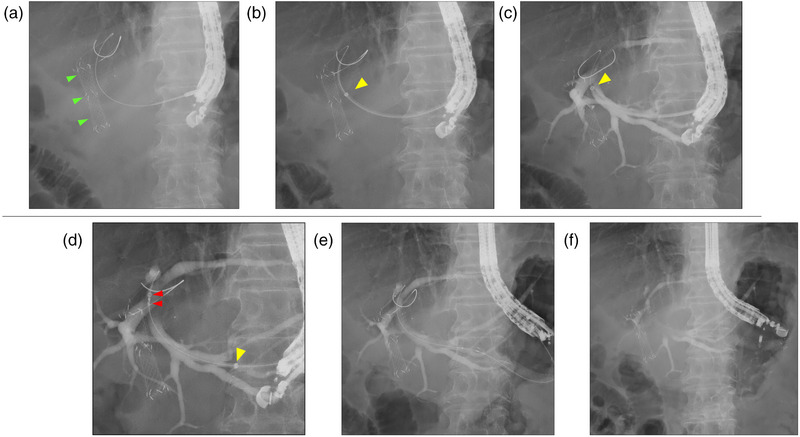
FIGURE 3.

Fluoroscopic cholangiogram showing the stenting technique using the tapered sheath dilator (EndoSheather) in endoscopic ultrasound‐guided hepaticogastrostomy (EUS‐HGS). Green arrowheads indicate the deployed stent under endoscopic retrograde cholangiopancreatography. (a) Proper insertion of a guidewire. (b) Utilization of the tapered sheath dilator to mechanically dilate the needle tract. Yellow arrowhead indicates the radiopaque marker of the outer sheath. (c) Removal of the inner catheter, leaving the outer sheath in position within the bile duct. The outer sheath enabled aspiration of bile juice and injection of contrast medium. (d–f) Smooth insertion and deployment of a fully covered self‐expandable metal stent with a 5.9 Fr delivery system. Red arrowheads show the markers of the metal stent.
