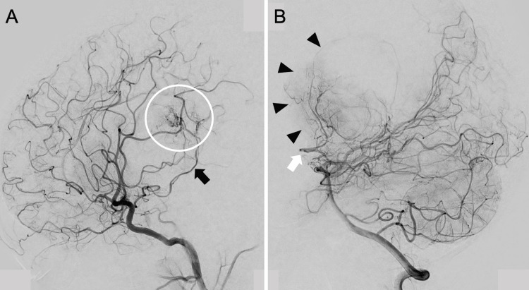
Figure 2. Preoperative angiogram showing multiple feeders and arterial-venous shunt in the tumor.
Right internal carotid angiogram showing that the feeder was the right anterior choroidal artery (black arrow) and arterial-venous shunt of the tumor (white circle) (A). Vertebral angiogram showing that the feeder was the right lateral posterior choroidal artery (white arrow) and tumor stain (black arrowheads) (B)

