Figure 5.
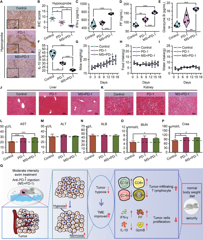
Exercise-Anti-PD-1 combined treatment alleviated TME hypoxia and improved the immune microenvironment with favorable biosafety. (A, B) Hypoxyprobe staining images (A) and IHC scores (B) of B16F10 tumor tissues in C57BL/6 mice with the specified treatments (scale bar: 100 μm, enlarged drawing: 50 μm; selected area: 5). (C–F) The detection of IFN-γ (C), Perforin (D) Granzyme B (E), and IL-10 (F) levels by ELISA in tumor tissues. (G–I) Weight (G) food intake (H) and water intake (I) of mice. (J, K) At the end of the experiment, using H&E to stain the liver (J) and kidney (K) from mice (scale bar: 100 μm). (L–N) Hepatotoxicity measured by aspartate aminotransferase (AST) (L), alanine aminotransferase (ALT) (M), and albumin (ALB) (N). (O, P) Nephrotoxicity measured by blood urea nitrogen (BUN) (O) and creatinine (Crea) (P). (Q) The schematic diagram of tumor reaction process after MS and Anti-PD-1 combination treatment. The data were presented as mean ± s.d. Statistical significance of the differences between groups was determined using t test. *, p<0.05; **, p<0.01; ***, p<0.001.

