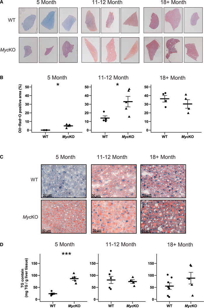
Figure 2. MycKO mice prematurely develop NAFLD.
(A) Representative oil red O (ORO)-stained liver sections of WT and MycKO mice.
(B) Quantification of ORO-stained sections. At least 3 liver sections from 4 or 5 mice were scanned, quantified, and combined.
(C) Higher-power magnification of the sections from (A) showing a greater prominence of large lipid droplets in MycKO livers.
(D) Triglyceride content of WT and MycKO livers. (B and C) Unpaired t test, *p < 0.05, ***p < 0.001. Error bars: standard deviation (SD).

