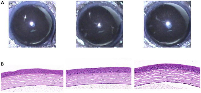
FIGURE 4.
Clinical and histological images of cornea in RCI001 group at week 5. (A) Anterior segment photographs of RCI001 group at week 5. There are no abnormal changes observed, such as corneal edema or opacity. (B) H&E staining of RCI001 group at week 5 shows healthy corneal epithelial findings and intact stromal and endothelial integrity. H&E, hematoxylin and eosin.

