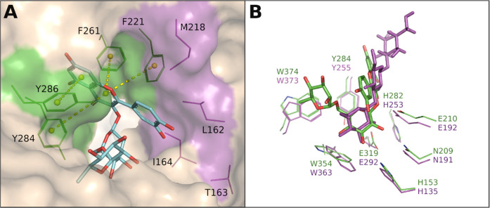
Figure 3.
Active site of AnRut with bound rutin. (A) Interactions between hydrophobic (magenta) and aromatic residues (green) in the +1 subsite of AnRut and docked rutin. The aromatic side chains of F221, F261, F284, and F286 clamp the aglycone moiety by π–π stacking interactions shown as yellow dotted lines.85,87 (B) Superposition of common residues in the −1 subsites of AnRut (carbon atoms in green, oxygen in red, and nitrogen in blue) and CaExg (magenta), depicting rutin (green-red) modeled into the active site of AnRut, and laminaritriose (β-d-Glc-(1 → 3)-β-d-Glc-(1 → 3)-Glc, magenta) cocrystallized with CaExg (PDBs 3N9K and 1EQC for the ligand and CaExg, respectively). The acid/base catalysts Glu210 and Glu192 are also shown.

